
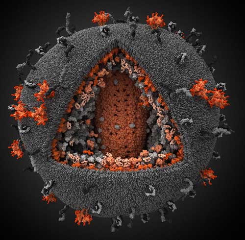
From the Russian company called Visual Science comes this absolutely stunning 3D visualization of the human immunodeficiency virus:
From the article on the Web site:
HIV virion is a roughly spherical particle with a diameter between 100 and 180 nm. Virion is surrounded by cell-derived lipid membrane containing surface proteins. Some of these proteins are products of viral genome (surface glycoprotein gp120/gp41) and others are captured from the host cell during viral budding (e.g. ICAM-1, HLA-DR1, CD55 and some others). The gp120/gp41 glycoprotein interacts with receptors on cell surface promoting fusion of virus and cell membranes. Other surface proteins found in HIV perform supporting functions. […]
The HIV genome is approximately 10000 nucleotides long and contains 9 genes, which encode 15 different proteins. The most important viral genes (open reading frames) are Gag, Pol and Env. Gag encodes the p55 protein, which is subsequently cut into structural proteins: MA, CA, NC and p6. Pol reading frame encodes integrase, protease, and reverse transcriptase. Env encodes the two subunits of the surface glycoprotein complex. Other genes (Tat, Rev, Vif, Vpr, Vpu and Nef) produce accessory proteins, which modulate host cell metabolism and facilitate different stages of HIV life cycle.
Click on the picture for a larger version and other visualizations showing different cross-sections of the virus.
It looks like it’s made of carpet! How cool is that. . .and doesn’t it look firey and dangerous too!
Wow
It looks like it’s made of carpet! How cool is that. . .and doesn’t it look firey and dangerous too!
Wow
I want someone to crochet this.
Perhaps these people could do it:
http://dreamzzzzzzzzzzzzzzz.blogspot.com/2009/09/hyperbolic-crochet-coral-reef.html
I want someone to crochet this.
Perhaps these people could do it:
http://dreamzzzzzzzzzzzzzzz.blogspot.com/2009/09/hyperbolic-crochet-coral-reef.html
I thought it was knitted.
I thought it was knitted.